Biofilms are formed when free-swimming, planktonic microbes adhere to one another and to a surface, and are often resistant to antibiotics. To test this antibiotic resistance, Emery Pharma offers a microplate peg lid-based Minimum Biofilm Eradication Concentration (MBEC) assay and crystal violet biomass staining assay in 96-well microplate format.
MBEC assays can be used to determine the minimum concentration of a test compound that will penetrate and eradicate microbial biofilms. This assay also allows for consistent biofilm generation and evaluation of multiple test compounds over a range of concentrations. Both antiseptic (as described in ASTM E2799-17) and antibiotic compounds can be tested.
Crystal violet is a dye that stains both microbial cells as well as extracellular matrix, and is commonly used as a readout for biomass. At Emery Pharma, crystal violet staining can be performed in conjunction with the MBEC assay or as an independent assay to evaluate biofilm biomass.

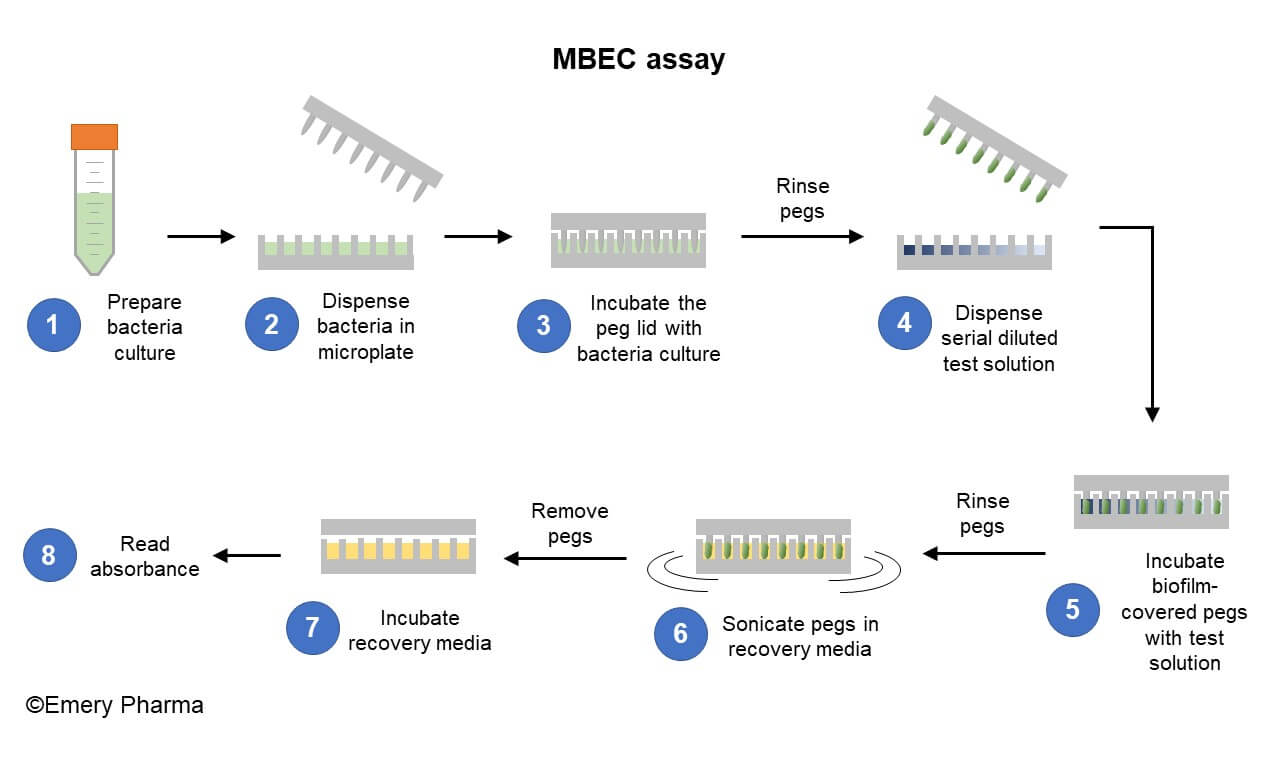
A step-by-step summary of the MBEC Assay. (1-2) Bacteria culture is prepared and dispensed into a 96-well microplate. (3) The peg lid is placed in the bacteria culture and incubated to generate the biofilm. (4) The peg lid is gently rinsed to removed planktonic bacteria and a serial diluted test solution is dispensed into a new 96-well microplate. (5) The pegs covered in biofilm are incubated in the test solutions. (6) The peg lid is again gently rinsed to remove planktonic bacteria and placed in a new 96-well microplate containing recovery media. The peg lid is then sonicated to dislodge the biofilm into the recovery media. (7) Following sonication, the peg lid is replaced with a regular 96-well microplate lid and the plate containing recovery media is incubated. (8) Following incubation, the OD650 absorbance is read on a spectrophotometer. Wells with an OD650 of less than 0.1 is evidence of biofilm eradication.

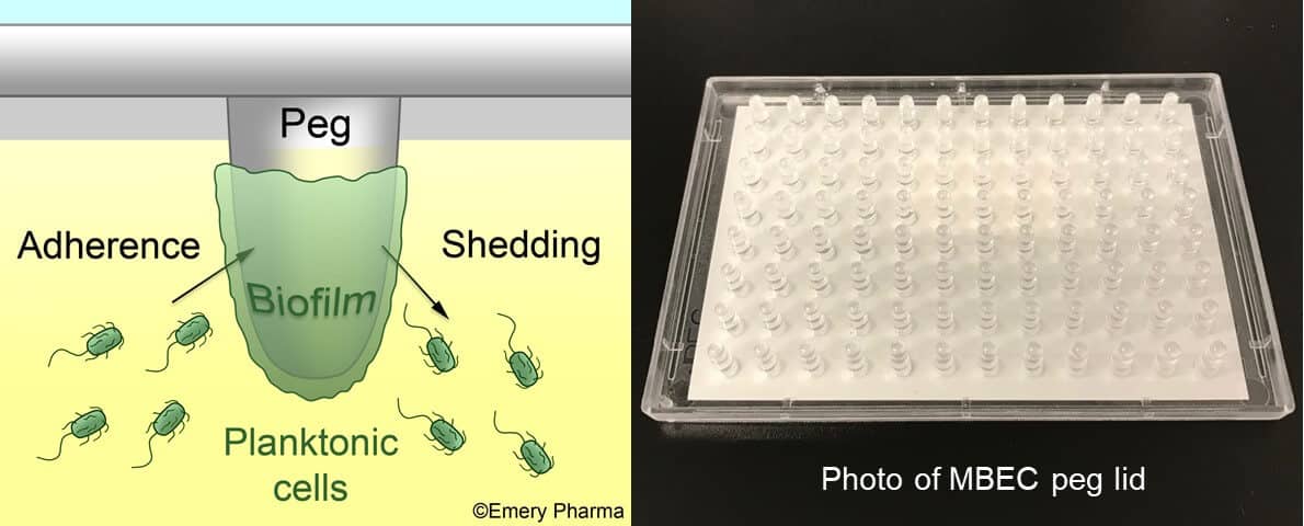
MBEC peg lid. (Left) Illustration depicting the adhesion of planktonic bacteria to the MBEC peg, the formation of the sessile biofilm, and the subsequent shedding of bacteria from the biofilm. (Right) A photograph of the MBEC peg lid.

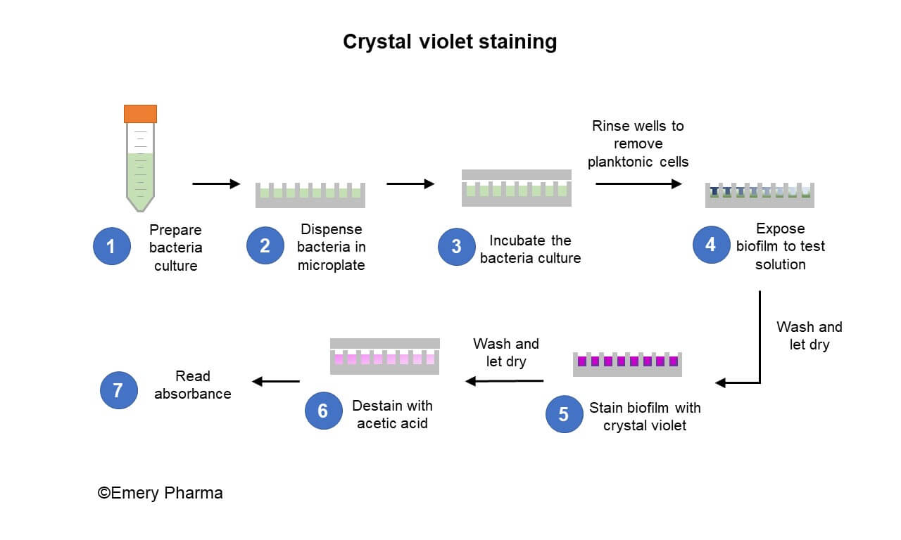
Crystal violet staining. (1-2) Bacteria culture is prepared and dispensed into a 96-well microplate. (3) The bacteria culture is incubated to generate the biofilm inside the wells. (4) The bacteria culture is then removed and the wells washed to remove planktonic cells before a serial diluted test solution is dispensed into the microplate. (5) After incubation of the biofilm with the test solution, the wells are washed, allowed to dry, then stained with crystal violet solution. (6) The stained biofilms are washed to remove excess dye and a volume of acetic acid solution is added to the wells to de-stain the crystal violet. (7) The absorbance of the de-stained acetic acid solution is read on a spectrophotometer.
Emery Pharma also offers custom biofilm eradication protocol development to suit the client’s needs. We can test various surfaces for biofilm formation and offer a vast inventory of over 1,500 isolates both bacterial clinical isolates and sample fungal strains available for screening, such as:
- Staphylococcus aureus
- Pseudomonas aeruginosa
- Candida albicans
- Candida parapsilosis
Reference
ASTM E2799-17, Standard Test Method for Testing Disinfectant Efficacy against Pseudomonas aeruginosa Biofilm using the MBEC Assay, ASTM International, West Conshohocken, PA, 2017